Functional Brain Mapping
Functional brain mapping is essential in neuroscience research and clinical applications to identify and understand specific brain regions' roles in various cognitive, sensory, and motor functions. This technique helps in assessing the brain's functional organization by identifying active areas during specific tasks or its response to stimuli. It is widely applied in areas such as pre-surgical planning, neurorehabilitation, and understanding neural connectivity and plasticity.
Setup Requirements and Technological Challenges
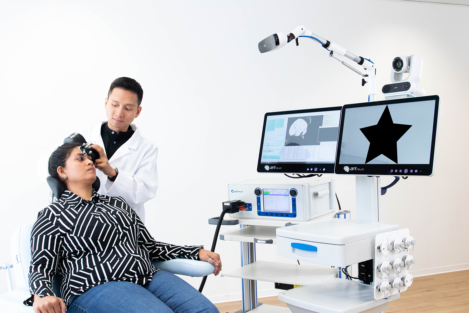
Functional brain mapping demands high spatial and temporal resolution to accurately capture rapid neural dynamics and localized brain activity. Non-invasive methods such as navigated TMS (nTMS), EEG, fMRI, MEG, and fNIRS systems are typically employed to record brain activity with high temporal and/or spatial precision. nTMS is also preferred as a method for localizing motor or linguistic function before surgical procedures as compared to fMRI due to its time-precision, high accuracy, non-invasiveness and low dependency on the subject. An nTMS session is not invasive and can be applied to most subject populations that do not cooperate with going under an MRI scanner.
Key challenges include:
- Achieving a balance between data quality and subject comfort
- Minimizing movement artefacts and noise in the data
- Effectively integrating multimodal data sources such as behavioral responses or imaging data.
- The use of precise and reliable optical tracking technology.
- Real-time visual feedback for TMS-EMG, for example, MEP responses.
- Export of mapped functional hotspots in popular formats such as DICOM for review in other softwares.

Our Solutions
To meet the demands of functional brain mapping, we provide a suite of solutions optimized for high-quality data acquisition, ease of use, and integration capabilities.
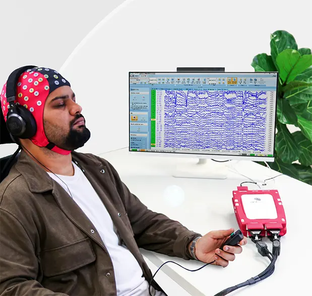
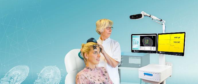
For nTMS purposes, the visor2™ system provides real-time navigation, accurate positioning for TMS applications and real-time visualization of TMS targets on subject specific brain images, allowing researchers and clinicians to map functional brain areas with high precision. The eego™mylab system also allows for high density EEG recordings at high sampling rates (up to 16384Hz) which is often preferred for TMS-EEG applications.
Showcases and Publications

Cortical Pathways During Postural Control: New Insights From Functional EEG Source Connectivity
F. Barollo et al.

The critical role of the inferior frontal cortex in establishing a prediction model for generating subsequent mismatch negativity (MMN): A TMS-EEG study
Lui, T. K. Y., Shum, Y. H., Xiao, X. Z., Wang, Y., Cheung, A. T. C., Chan, S. S. M., Neggers, S. F. W., & Tse, C. Y.

Stimulation of the left dorsolateral prefrontal cortex with slow rTMS enhances verbal memory formation
Van der Plas M, Braun V, Stauch BJ, Hanslmayr S.

Sponge EEG is equivalent regarding signal quality, but faster than routine EEG
Michael Günther, Leonie Schuster, Christian Boßelmann, Holger Lerche, Ulf Ziemann, Katharina Feil, Justus Marquetand.


