- Home
- About ANT
-
Products
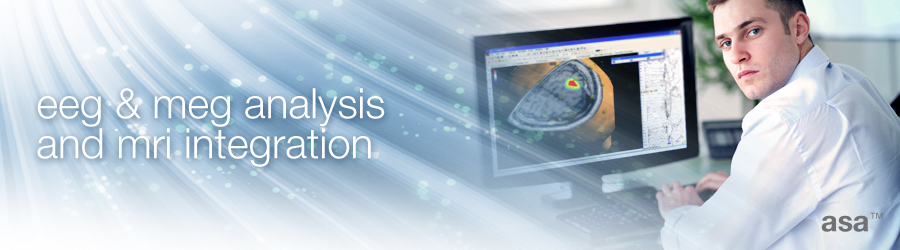
asa
asa is a highly flexible EEG/ERP and MEG analysis package with a variety of source reconstruction, signal analysis and MRI processing features.
.jpg)
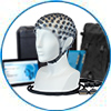
eego mylab
The new frontier in multimodal brain research. With up to 16 kHz sampling rate, 256 EEG channels and unique software features, eego mylab gives you an unprecedented in-depth understanding of the human brain.
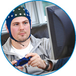
eego sports
eego sports offers complete freedom to collect high-density EEG data, bipolar EMG signals, and a variety of physiological sensor data, wherever and whenever required, with publish quality data in less than 15 minutes!
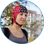
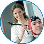
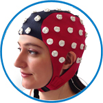
waveguard net
The waveguard net sets a new standard for research applications requiring high-density EEG data acquisition with quick preparation time, high flexibility, and subject comfort.
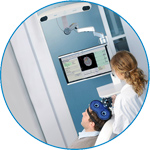
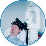
visor2
Our new and upgraded visor2 solutions integrate all the latest technologies for navigated rTMS, dual-coil navigation support, EEG-TMS recordings and pre-surgical evaluation for the highest quality in research and clinical procedures.
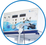
powerMAG ANT
The PowerMAG ANT 100 rTMS stimulator is designed for the specific needs of high-end TMS applications. Powerful high-frequency TMS as well as high precise single pulse and repetitive pulse protocols are combined in one single device.
xensor
xensor offers the solution for digitization of 3D electrode positions. xensor takes care of the whole procedure; it records, visualizes and stores positions acquired with a dedicated digitizer.
waveguard original
waveguard original is the cap solution for EEG measurements compatible with fMRI, MEG and TMS system. Use of active shielding guarantees performance in even the most demanding environments.
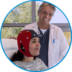
waveguard connect
waveguard connect EEG caps are a perfect match for hospitals and institutes aiming at reliable EEG, maximum uptime and great patient comfort! For optimal signal quality, the electrodes are made of pure, solid tin.
waveguard touch
waveguard touch is a dry electrode EEG cap. The unique Ag/AgCl coated soft polymer electrodes provide stable, research-grade EEG signals while maintaining subject comfort. The combination of these innovative dry electrodes and the industry-leading waveguard cap makes waveguard touch the best solution for dry EEG.
smartmove
smartmove allows planning of a complete TMS session ahead by defining stimulation sites based on anatomical MRI information and functional information like fMRI, PET or EEG/MEG.
- References
- Support
- Events
- News
- Contact Us
 Read more
.jpg)
You are here
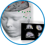
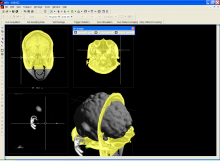
MRI and head modeling
MRI and head modeling
asa uses the latest technology available for rendering of pixelated images and 3D objects with optimal performance.
The MRI view allows the free rotation of slices cut through the MRI. It combines the display of MRI, segmentation of head compartments, inverse reconstructions (such as MUSIC, LORETA, cortical imaging and cortical ERD/ERS, and dipole fit results), and fMRI.
In addition, all objects usually shown in the 3D views (such as EEG/MEG maps, head models and sensors) can be added to the display.
Talairach Coordinate System and Transformations
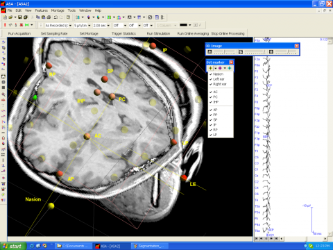 asa allows to transform results obtained from an individual into a standardized coordinate system based on the Talairach definition. For this purpose, a set of anatomical landmarks is placed on the subject's MRI, based on which the transformation is carried out.
The transformed MRI (which is quite different from the individual MRI, since orientation and spacing between MR slices are newly defined and applied to create a new block of MRI) is then exported in to asa or Analyze (SPM) formats.
The same export can be applied to LORETA and MUSIC results in order to obtain results that can be further processed in a group statistics.
 Segmentation of MRI and CT


ANT Neuro
Welbergweg 74
7556 PE Hengelo
Netherlands
T: +31 (0) 85 049 8175
F: +31 (0) 85 049 3919
E: Send us an email