- Home
- About ANT
-
Products
asa
asa is a highly flexible EEG/ERP and MEG analysis package with a variety of source reconstruction, signal analysis and MRI processing features.
.jpg)
eego mylab
The new frontier in multimodal brain research. With up to 16 kHz sampling rate, 256 EEG channels and unique software features, eego mylab gives you an unprecedented in-depth understanding of the human brain.
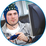
eego sports
eego sports offers complete freedom to collect high-density EEG data, bipolar EMG signals, and a variety of physiological sensor data, wherever and whenever required, with publish quality data in less than 15 minutes!
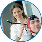
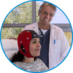
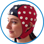
waveguard net
The waveguard net sets a new standard for research applications requiring high-density EEG data acquisition with quick preparation time, high flexibility, and subject comfort.
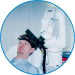
visor2
Our new and upgraded visor2 solutions integrate all the latest technologies for navigated rTMS, dual-coil navigation support, EEG-TMS recordings and pre-surgical evaluation for the highest quality in research and clinical procedures.
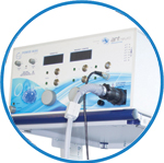
powerMAG ANT
The PowerMAG ANT 100 rTMS stimulator is designed for the specific needs of high-end TMS applications. Powerful high-frequency TMS as well as high precise single pulse and repetitive pulse protocols are combined in one single device.
xensor
xensor offers the solution for digitization of 3D electrode positions. xensor takes care of the whole procedure; it records, visualizes and stores positions acquired with a dedicated digitizer.
waveguard original
waveguard original is the cap solution for EEG measurements compatible with fMRI, MEG and TMS system. Use of active shielding guarantees performance in even the most demanding environments.
waveguard connect
waveguard connect EEG caps are a perfect match for hospitals and institutes aiming at reliable EEG, maximum uptime and great patient comfort! For optimal signal quality, the electrodes are made of pure, solid tin.
waveguard touch
waveguard touch is a dry electrode EEG cap. The unique Ag/AgCl coated soft polymer electrodes provide stable, research-grade EEG signals while maintaining subject comfort. The combination of these innovative dry electrodes and the industry-leading waveguard cap makes waveguard touch the best solution for dry EEG.
smartmove
smartmove allows planning of a complete TMS session ahead by defining stimulation sites based on anatomical MRI information and functional information like fMRI, PET or EEG/MEG.
- References
- Support
- Events
- News
- Contact Us
 Read more
.jpg)

You are here
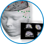
High resolution EEG and source localization in neonates
High resolution EEG and source localization in neonates
Although Electroencephalography (EEG) source localization is being widely used in adults, this promising technique has not yet been applied to newborns because of technical difficulties, such as lack of data concerning the newborn skull conductivity, thickness, and homogeneity. Using a new type of EEG headcap molded on each baby's head, we aimed to determine whether this technique could be adapted to neonates, and to evaluate the importance of these technical difficulties. We carried out EEG source reconstruction of the recordings of five neonates using dipole fit algorithm. We used four different head models for each neonate, obtained from individual MRI scans: normal skull thickness and conductivity of 0.0042 S/m; normal thickness and conductivity of 0.33 S/m; increased thickness and conductivity of 0.0042 S/m; and normal thickness and conductivity with a modeled bregma fontanel. Dipole locations were consistent with MRI and clinical data. The mean difference between the dipole locations in the 0.0042 and the 0.33 S/m skull layer models was 11.6 ± 2.5 mm, with an average 29.7% decrease in magnitude for the 0.33 S/m model but no significant changes for the dipoles orientation. Skull layer thickness had a large influence on magnitude, but no significant effect on position and orientation. The mean difference between the dipole locations induced by the modeled fontanel was 2.0 ± 2.1 mm, with an average 2.1% increase in magnitude. Our results show that EEG source localization is feasible in neonates. With further development, the technique may prove useful for neurological evaluation of neonates.


ANT Neuro
Welbergweg 74
7556 PE Hengelo
Netherlands
T: +31 (0) 85 049 8175
F: +31 (0) 85 049 3919
E: Send us an email